
A study of cervical length measured ultrasonographically in prediction of preterm delivery
Abstract
Introduction- Cervical length appears to be an efficient test for predicting preterm birth. Transvaginal sonography (TVS) is the preferred route for cervical assessment to identify women at increased risk of spontaneous preterm birth and may be offered to women at increased risk of preterm birth.
Methods- This was prospective observational study conducted in Obstetrics and Gynecology department of SMS Medical college, Jaipur, Rajasthan, Indiafrom August 2015 to July 2016. Out of obstetric cases attending antenatal OPD, cases of singleton pregnancies were selected at random. In this study all the participants were divided into 2 groups: Each group include 100 patients. Every participant underwent a transvaginal sonography (TVS), using probe of 5 to 7.5 MHz, measuring cervical length.
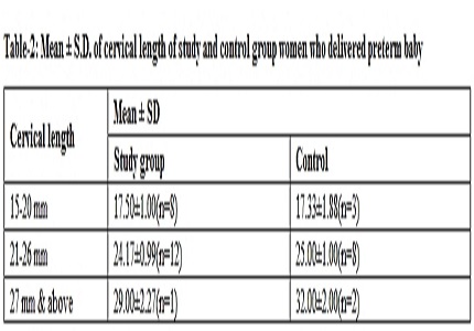
Results- About 39 women in control group and 36 women in study group were primigravida, remaining were multigravida. History of preterm labour was present in 27 women in study group and 28 women in control group. Cervical length measurement was 21-26 mm in 30 (30%) women and among them 12 (44.44%) delivered preterm. Mean birth weight was 1.75±0.04 in control group and 1.75±0.06 in study group in which cervical length was between 21-26 mm. In our study in study group revealed Prevalence – 27%, Positive predictive value – 52.10%, Negative predictive value – 88.70%, Sensitivity – 81.48%, Specificity – 75.34%.
Conclusion- We found that TVS had good sensitivity, specificity, predictive value in both group. Thus measurement of cervical length by TVS can be used to predict increase risk of preterm delivery cases with threatened preterm labor.
Downloads
References
2. Kurpa FG, Faltin D, Cecatti JG, Surita FGC, Souza JP. Predictors of preterm birth. Int J Gynecol Obstet. 2006;94:5-11. [PubMed]
3. Yoshizato T, Obama H, Nojiri T, Miyaka.Y, Miyamoto S, Kawarabayashi T. Clinical significance of cervical length shortening before 31 week’s gestation assessed by longitudinal observation using transvaginal ultrasonography. J Obstet Gynaecol. Res. 2008;34:805-11. [PubMed]
4. Masamoto H, Nagai Y, Inamine M. et al. Outcome of pregnancy after laser conization: implications for infection as a causal link with preterm birth. J ObstetGynaecol Res. 2008;34:838-842. [PubMed]
5. Lim K, Butt K, Crane JM.Diagnostic imaging committee; family physicians advisory committee; maternal fetal medicine committee. SOGC Clinical Practice Guideline. Ultrasonographiccervicallengthassessment in predictingpreterm birth in singletonpregnancies. J ObstetGynaecol Can. 2011 May;33(5):486-499. doi: 10.1016/S1701-2163(16)34884-8. [PubMed]
6. Melamed N, Hiersch L, Domniz N, Maresky A, Bardin R, Yogev Y. Predictive value of cervical length in women with threatened preterm labor. Obstet Gynecol. 2013 Dec;122(6):1279-87. doi: 10.1097/AOG.0000000000000022. [PubMed]
7. Rozenberg P, Goffinet F, Kayem G, Perdu M, Phillipoe HJ, Misand J. The value of intravaginal ultrasonography of the cervix uteri for evaluation of the risk of premature labour. Jr de Gynecologic Obstetrics et biologic de la Reproduction.1997;26(6):623-29.
8. Kore SJ, Parikh MP, Lakhotia, Kulkarni, Ambiye VR. Prediction of risk of preterm delivery by cervical assessment by transvaginal ultrasonography. J ObstetGynecol India. 2009;59(2):131-35.
9. Qudah S, Athamneh T, Tawlbeh A, Daklallah L, Al- hajji M. Cervical length as a predictor risk of preterm delivery. Journal of Royal Medical services. 2017 March;24(1):18-21.
10. Khusboo, K Dinesh, Verma A, Chaursia S, Nag R. Cervical sonomorphometric evaluation of normal and preterm labour by transvaginal and transabdominal sonography. Int J Reprod Contracept Obstet gynecol.2017 Feb;6(2):417-422.
11. Begum J, Behera AK. Cervical length by ultrasound as a predictor of preterm labour. Int J Reprod Contracept Obstet gynecol.2014 Sept;3(3):646-652.
12. Iams JD, Goldenberg RL, Meis PJ, Mercer BM, Moawad A, Das A, Thom E, McNellis D, Copper RL, Johnson F, Roberts JM. The length of the cervix and the risk ofspontaneouspremature delivery. National Institute of Child Health and Human Development Maternal Fetal Medicine Unit Network. NEngl J Med. 1996 Feb 29;334(9):567-72. [PubMed]
13. Mukherji J, Anant M, Ghosh S, Bhattacharyya SK, Hazra A, Kamilya GS. Normative data of cervical length in singleton pregnancy in women attending a tertiary care hospital in eastern India. Indian J Med Res. 2011;133(5):492-6.
14. Berghella V, TolosaJE,Kuhlman K, Welner S, Bolognese RJ,Wepner RJ. Cervical ultrasonography compared with manual examination as predictor of preterm delivery. Am. J. Obstetrics and gynaecology. 1997 Oct.;177(4):723-30. [PubMed]
15. Wadhawan UT, Shah NP, Patil AN. Prediction of cervical length by cervical length. Int J Reprod Contracept Obstet gynecol.2017 July;6(7):2978-2982.
16. Crane JM, Van den Hof M, Armson BA, Liston R. Transvaginalultrasound in the prediction of preterm delivery: singleton and twingestations.Obstet Gynecol. 1997 Sep;90(3):357-63. [PubMed]
Copyright (c) 2017 Author (s). Published by Siddharth Health Research and Social Welfare Society

This work is licensed under a Creative Commons Attribution 4.0 International License.

 OAI - Open Archives Initiative


