
Cardiotocographic evaluation of fetal condition and outcome in pregnant women presenting with lessfoetal movement beyond 34 weeks
Abstract
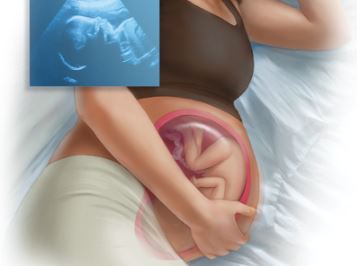
Introduction: Cardiotocography (CTG) is widely used to identify pregnancies that might be benefited from continuous fetal monitoring in labour or during antepartum states. The CTG is a valuable tool for early detection of fetal compromise. It had the modest predictive value for soft outcomes such as mild asphyxia, fetal distress and increased operative delivery particularly in the antepartum and intrapartum condition. So the objective of the preset study is to observe the fetal condition and outcome in pregnant women presenting with less foetal movement beyond 34 weeks. Methods: For that 50 cases of pregnant women beyond 34 weeks presenting with less fetal movement were included and CTG was done. They were observed till delivery and APGAR score of the neonate were collected at 1st minute and at 5th minute. When CTG was found reactive before 37 weeks they were managed conservatively and again examined at 37 weeks. When CTG was found nonreactive after 37 weeks, they were reevaluated after 30 minutes and decision was taken. Data were correlated and analyzed by SPSS 20.Results: Different sociodemographic profiles where majority of women were at age group 21-25 years, working as housewives, nonsmokers and found to be from rural areas with lower and lower middleclass status. Average gestational age was found 38 weeks, mean height of uterus was 36 cm. Maximum patient were primigravida and with no medical illness. 100% patients were presented with vertex presentation. Cardiotocographic findings revealed baseline fetal heart rate was found 110 to 160 bpm in 34(68%) cases and < 110, > 160 bpm found in 16(32%) cases. Also baseline variability, acceleration and decelerations were found. Among 50 cases 34(68%) had normal vaginal delivery and 16(32%) cases undergone CS. Neonatal outcome revealed live birth was 50(100%), <5 APGAR score was found in 5(10%), 5(10%) cases required neonatal resuscitation, 5(10%) needed NICU admission. No seizures within 24-48 hours and no cases of perinatal death. Conclusion: Cardiotocography is an important tool to assess the fetal condition in pregnancy.
Downloads
References
2. Amnon Hader, Eyal Sheiner. Abnormal Fetal Heart Rate Pattern During First Stage OfLabour And Effect On Perinatal Outcome, American Journal of Obstetrics And Gynecology, 2001;185(4):863-868, october 2001.
3. Odomgo BE, Ndavi PM. Cardiotocography And Perinatal Outcome In Women With And Without Meconium Stained Liquour, East Afr Med J, 2010;87(5):199-204.
4. Eugenia Maria. Low APGAR Score At 5 Minutes In A Low Risk Population; Maternal And Obstetrics Factors And Perinatal Outcome, Rev Assoc. Med Bras. 2012; 58:12- 19.
5. Eugene Bailey Intrapartum Fetal Monitoring, Am Fam Physician 2009;15;80 (12):1388-1396
6. Alfirevic Z, Devane D, Gyte GM. Continuous CTG As A Form Of Electronic Fetal Monitoring For Fetal Assessment During Labour, Conhrane Database of Systematic Review 2006 ;3:54-56.
7. Low JA, Pickersgill H. Prediction And Prevention Of Intrapartum Fetal Asphyxia In Term Pregnancies, .American.Medica Journal,. Obstet Gynecol. 2001;184(4): 724- 730
8. Rozen A,Sheiner E. Abnormal Fetal Heart Rate Trecings And Congenital Fetal Hypothyroidism, Int J. Fertil Womens Med 2008;51(6): 267-269
9. Fawole AO, Sotiloye OS, Oladimeji AO, Alao MO, Hunyinbo KI, Sadoh EA, Otolorin EO. Antenatal cardiotocography: experience in a Nigerian tertiary hospital. Nigerian Postgraduate Medical Journal 2008; 15:19-23.
10. Mires G, Williams F, Howie P. Randomised controlled trial of cardiotocography versus Doppler auscultation of the fetal heart at admission in labour in low-risk obstetric population. BMJ 2001; 322: 1457–62.
Copyright (c) 2025 Author (s). Published by Siddharth Health Research and Social Welfare Society

This work is licensed under a Creative Commons Attribution 4.0 International License.

 OAI - Open Archives Initiative


