E-ISSN:2455-5444
P-ISSN:2581-4389
RNI:MPENG/2017/74037

Research Article

Severe Preeclampsia

Obs Gyne Review - Journal of Obstetric and Gynecology

2024 Volume 10 Number 1 Jan-Dec
Publisherwww.medresearch.in

Sultana S1*, Siddique MAB2, Hoque S3, Shahriar SM4, Karim K5
DOI:https://doi.org/10.17511/joog.2024.i01.01

1* Salma Sultana, Junior Consultant, Obst and Gaynae, 250 Bedded General Hospital, Thakurgaon, Bangladesh.

2 Md Abu Baker Siddique, Assistant Professor, MS Orthopaedic, M Abdur Rahim Medical College, Dinajpur, Bangladesh.

3 Smrity Hoque, Junior Consultant, Obst and Gaynae, Current Charge Baliadangi Upazila Health Complex, Thakurgaon, Bangladesh.

4 Shihab Mahmud Shahriar, Junior Consultant, Surgery, 250 Bedded General Hospital, Thakurgaon, Bangladesh.

5 Khadiza Karim, Medical Officer, Obst and Gaynae, 250 Bedded General Hospital, Thakurgaon, Bangladesh.

Background: Hypertensive disorders represent the most common medical complication of pregnancy affecting between 7 to 15 percent of all gestation and account for approximately a quarter of all antenatal admission. In Bangladesh, the incidence of this killer disease is still high and it is the third major cause of maternal death in our country.

Objective: To evaluate pregnancy outcomes in severe preeclampsia.

Methods: The data were collected through the active participation of the patients' interviews by the performed proforma of data collection sheet and then data were gathered, decorated, and tabulated after data cleaning and edition. Then the results were found and they were tested by the Student's unpaired t-test (quantitative data) and chi-square test (qualitative data) to see their level of significance i,e p-value which was set as the cutoff level at <0.05. So if the p-value is >0.05 the results are not significant.

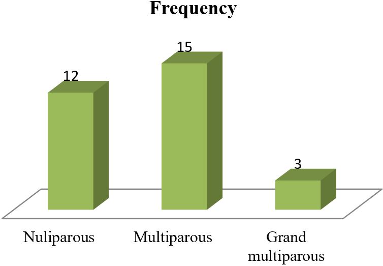
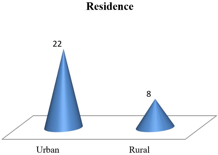
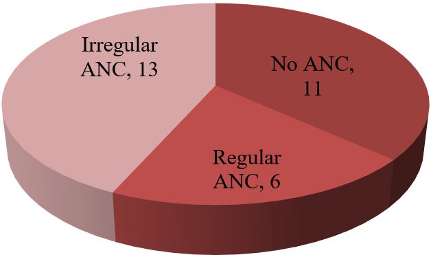
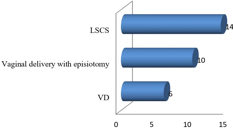
Results: The data analysis of 30 patients yielded the following results. The mean age of 30 mothers was 27.65 (± 5.85) years. The maximum 19 (63.33%) patients were from the 20-34 years age group. Among the 30 mothers with severe preeclampsia, 12(40%) were primiparous and the rest 18 (60%) were multiparous. To ensure safe delivery 14(46.67%) mothers adopted LSCS whereas 10(33.33%) adopted vaginal delivery with episiotomy. The rest 6 (20%) underwent VD. Regular ANC ensured only 6 (20%) respondents whereas the rest 80% replied that they were either irregular ANC or no ANC. The most common complications mothers faced were abruptio placenta (28.57%) followed by convulsion (14.28%) and HELLP syndrome (9.52%). Among the babies, 5% showed APGAR score above 7 at 1st minute whereas 58% showed the same score at 5 minutes.

Conclusion: In conclusion, it may be assumed that though various fetal and maternal morbidities may occur in the case of severe preeclampsia mortality is seen only in the case of neonates. Maternal mortality has almost disappeared from our perspective in the case of severe pre-eclampsia.

Keywords: Sultana S et al. Evaluation of Pregnancy Outcome in Severe Preeclampsia

Corresponding Author How to Cite this Article To Browse
Salma Sultana, Junior Consultant, Obst and Gaynae, 250 Bedded General Hospital, Thakurgaon, , Bangladesh.
Email:
Sultana S, Siddique MAB, Hoque S, Shahriar SM, Karim K, . Obs Gyne Review J Obstet Gynecol. 2024;10(1):9-15.
Available From
https://obstetrics.medresearch.in/index.php/joog/article/view/168

Manuscript Received Review Round 1 Review Round 2 Review Round 3 Accepted
2024-08-01 2024-08-07 2024-08-13 2024-08-19 2024-08-25
Conflict of Interest Funding Ethical Approval Plagiarism X-checker Note
None Nil Yes 11.32

© 2024by Sultana S, Siddique MAB, Hoque S, Shahriar SM, Karim Kand Published by Siddharth Health Research and Social Welfare Society. This is an Open Access article licensed under a Creative Commons Attribution 4.0 International License https://creativecommons.org/licenses/by/4.0/ unported [CC BY 4.0].

Introduction

Pre-eclampsia is a series of multi-system abnormalities usually occurring after 20 weeks of gestation with an ensuring diagnosis of new-onset hypertension (blood pressure at or above 140/90 mmHg) on two occasions at least 4 hours apart with significant proteinuria (a finding of protein in urine > 300 mg over a 24 hrs period) in a previous normotensive and nonproteinuric patient. Globally, each year more than four million women develop preeclampsia and approximately 100,000 women will have an eclamptic convulsion with over 90% occurring in developing countries[1].

It results in 12% of maternal deaths globally, up to 40% of maternal deaths in some countries and is responsible for the occurrence of up to 13% of stillbirths and 20% of early neonatal deaths [2]. Approximately 10-15% of maternal deaths in developing countries are associated with preeclampsia leading to eclampsia[3].

There is no concrete data found on the incidence of preeclampsia in our country, but calculated from the US Census Bureau, International Data Base, 2004, the extrapolated annual incidence of preeclampsia in Bangladesh is 76,032[4]. Our neighbouring country, India has incidence of preeclampsia, as recorded from hospital statistics, varying widely from 5 to 15% [5].

Bangladesh, the most densely populated country in South East Asia has high maternal mortality as well as perinatal mortality rate [6]. According to WHO severe preeclampsia accounts for 16.1% of maternal deaths in developed countries over the past 2 decades [7].

In pre-eclampsia, the normal endovascular invasion of cytotrophoblast into the spiral arteries fails to occur beyond the decidua-myometrial junction. As a result, the musculoelastic media in the myometrial segment remains responsive to vasoconstrictor stimuli resulting in decreased blood flow. There is acute atherosis of spiral arteries with obliteration of the lumen. Maternal illness varies from mild asymptomatic hypertension to neurological, renal, and cardiopulmonary compromise.

Concomitantly, some fetuses are healthy, while others experience severe intrauterine growth restriction. The maternal and perinatal mortality is thought to be <1% and 1% to 2% respectively.

While preeclampsia has the potential for serious complications, most cases of preeclampsia are mild and require minimal clinical treatment like increased maternal and fetal surveillance, blood pressure control and seizure prophylaxis, but ultimately delivery of the infant is the definitive treatment.

The decision to transfer women with preeclampsia for management and delivery elsewhere can create social hardship and may introduce morbidity risk for the infant. The challenge to clinicians lies in identifying patients who will suffer subsequent adverse outcomes from preeclampsia to intervene appropriately while minimizing unnecessary and potentially harmful interventions in patients who do not require them.

Severe preeclampsia and eclampsia are not distinct disorders but are differentiated according to their clinical syndrome. Although seizures define the condition of eclampsia, other neurological findings may be observed earlier in the continuum of preeclampsia. In preeclampsia, hypertension and proteinuria are present and when convulsions occur in addition to these signs, the condition is referred to as eclampsia [8]. The main aim of this study was to evaluate pregnancy outcomes in severe preeclampsia.

Materials and Methods

Study design: Observational cross-sectional study.

Place of study: The study was conducted among the admitted patients in the Department of Obstetrics &Gynaecology at BSMMU, Dhaka, Bangladesh.

Study population: The study was conducted among patients with severe preeclampsia admitted to the Department of Obstetrics & Gynaecology, BSMMU, Bangladesh.

Period of study: March 2015 to August 2015.

Sample size: 30 patients were selected by purposive sampling.

Inclusion Criteria

  • Pregnant women between 20-40 weeks of gestation.
  • Blood pressure ≥160/110mm of Hg with proteinuria.
  • Single intrauterine pregnancy.

Exclusion Criteria

  • Pregnant women <20 weeks and >40 weeks of gestation.
  • Gestational Hypertension.
  • Chronic Hypertension.
  • Antepartum Hemorrhage.
  • Presence of Diabetes mellitus.
  • Heart diseases.
  • Renal diseases.
  • HELLP syndrome.
  • Babies with Congenital Malformations.

Methodology: The interviews of the mothers were taken through a pre-structured questionnaire. The mothers were clinically assessed and supporting investigations were done to confirm the presence of medical and obstetrical risk factors contributing to severe preeclampsia. All the data were gathered, edited, decorated and finalized in figure and table form. As there was no hypothesis to be tested no statistical test was performed. The table and figure were plotted according to age, educational status, socio-economic status, BMI, mode of delivery, medical and obstetrical risk factors, APGAR score, perinatal outcome, neonatal death and their interval. The individual categorizations are given on the next page as the observations and results of this study. As the total sample size is 30 in this study the number of patients also manifests the percentage of patients here.

Main outcome variables studied: Preeclampsia- maternal outcome, perinatal age, parity, severity, mode of delivery, maternal complications and maternal mortality, perinatal outcome, birth weight, APGAR score, NICU admission, perinatal complications and perinatal mortality.

Procedures of data analysis and interpretation: After collection, data editing and clearing were done manually and prepared for data entry and analysis by using SPSS version 17. Student t-tests were done to compare the groups. P values <0.05 will be considered as significant.

Results

Out of 30 patients, the maximum number of patients were from the age group 20-34 years category.

The number was 19 (63.33%). Out of 30 mothers, 14 were nulliparous which was about 46.67% of the total study population.

The minimum number of pre-eclamptic mothers was grand multiparous (table-1& fig-I).

Table 1: Distribution of age and Mean age (n=30)

Age categoryNo. of mothersPercentage of mothers
<20 years620%
20-34 years1963.33%
≥ 35 years516.67%
Total30100%
Mean age ± SD (years)27.65±5.85

obstetrics_168_01.jpg
Figure 1:
Distribution of parity (n=30)

Out of 30 respondents of this study 22(73.33%) were urban residents and 8(26.67%) were rural residents (fig-II).

obstetrics_168_02.jpg
Figure 2:
Distribution of residence (n=30)

Maximum mothers in this study did not undergo regular antenatal check-ups. The real statistics are shown below (fig-III).


obstetrics_168_03.jpg
Figure 3:
Distribution of mothers receiving antenatal check-ups (n=30)

Table 2: Distribution of CNS manifestation (n=30)

CNS ManifestationNo. of Pre-eclamptic mothersPercentage of pre-eclamptic mothers
Headache1756.67%
Headache & Dizziness516.66%
Headache & Visual disturbances723.33%
Dizziness13.33%

Headache was the chief Central Nervous System manifestation found in this study. 17(56.67%) patients out of 30 were affected by headache (table-2).

Table 3: Distribution of GIT symptoms (n=30)

GIT symptomsNo. of pre-eclamptic mothersPercentage of pre-eclamptic mothers
Epigastric pain1343.33%
Vomiting and epigastric pain723.33%
Vomiting620%
Vomiting and nausea310%
Nausea13.33%
Total30100%

13 (43.33%) patients manifested epigastric pain as a Gastrointestinal symptom which was the maximum figure among 30 patients (table-3).

Table 4: Distribution of maternal complications

Maternal complicationsNumber %
Abruptio placenta6 (28.57%)
Convulsion3 (14.28%)
PPH3 (10.00%)
HELLP syndrome2 (9.52%)
Pulmonary edema2 (9.52%)
Renal failure1 (4.76%)

Out of 30 patients the maximum number of mothers had caesarean section. The rest number of patients underwent vaginal delivery (fig-IV).

obstetrics_168_04.jpg
Figure 4:
Distribution of mode of delivery (n=30)

Out of 30 severe preeclamptic mothers, complications were observed in 17 patients (table-4).

Table 5: Distribution by fetal outcome (n=30)

1.  APAGAR Score at 1 minute
7-104-61-30
5%87%5%5%
2.  APAGAR Score at 5 minute
7-104-61-30
58%40%2%5%
3.  Resuscitation (needed) 62%
4.  Admission into neonatal ward 45%

At one minute after birth, the maximum neonates (87%) showed the APGAR SCORE 4-6 and after 5 minutes (58%) neonates showed the APGAR SCORE 7-10. 62(%) newborn required resuscitation and 45(%) required admission into the neonatal ward (table-5).

Table 6: Distribution of newborn according to their birth weight (n=30).

WLNo of newborn
<2.5 kg18 (60%)
2.5-3 kg12 (40%)

Most of the newborn 18 (60%) were low birth weight babies (table-6).

Table 7: Distribution of neonatal mortality

WeightMortalityPercentage
<2.5 kg316.67
2.5 to 3 kg18.33

Out of 30 neonates, 4 babies died after birth. Among them, 3 (16.67%) were low birth weight babies (table-7).


Table 8: Distribution of complications in neonates

Problem in infants% of mothers (approx.)
Low birth weight (LBW)60%
Intrauterine growth retardation (IUGR)18.67%
Birth asphyxia16%
Hyper bilirubinemia4%
Neonatal septicemia2.33%

Out of 30 neonates, 60% were born with low birth weight (table-8).

Discussion

This observational cross-sectional study was conducted in the Department of Obstetrics and Gynaecology, BSMMU during 6 months to evaluate pregnancy outcomes of severe preeclampsia. For this purpose, 30 pregnant women were purposively selected according to inclusion and exclusion criteria.

This study showed maximum patients (63.30%) were age group of 20 to 34 years and the mean age of the total study population was 27.65±5.85years. These figures indicate that younger women were more affected by severe preeclampsia. This corresponds with the studies of Moodley and Mashioane where the mean age was 28 years and also similar to Brown MA and Buddle ML [9].

Half of the severe pre-eclamptic women in this study were multiparous. Conde-Aguedelo and Belizam JM also found a similar pattern of risk factors among nulliparous and multiparous women [10]. Among the 17 (56.67%) multiparous women 13 (76.47%) had pregnancy-induced hypertension in their previous pregnancy. The presence of gestational hypertension in a previous pregnancy is a known risk factor for gestational hypertension in a subsequent pregnancy [11].

In my study 22 (73.33%) were urban residents among them 15 patients came from low-income families with poor nutritional status. The maximum number of mothers in this study did not undergo regular antenatal check-ups. The cause behind this unwanted scenario revealed a lack of knowledge, low socio-economic condition, lack of awareness, ignorance of family and lack of knowledge about the health facilities around them. Patients with severe eclampsia present with various symptoms but the commonest were headache, headache with visual disturbance, epigastric pain, and epigastric pain with vomiting.

The mode of delivery was chosen caesarean section in the case of 14 (46.67%) severe preeclampsia mothers for better perinatal outcomes whereas 10(33.33%) mothers delivered vaginally with episiotomy and 6(20%) delivered vaginally. The results were consistent with a report presented by Nabila et al. [12].

Abruptio placenta was observed as the maximal maternal complication (28.58%) in this study. This result is similar to that of Lee and his colleagues' report where 32% of the pre-eclamptic women had the same maternal complication [13]. The commonest complications in this study were abruptio placenta, convulsion and HELLP syndrome. These complications were similar to the findings of Murphy DJ and Stirrat GM [14] Abruptio placenta was the commonest maternal complication in our study which was also observed in the study of Al-Mulhim and his colleagues [15].

Regarding APGAR score this study found that 95% of neonates achieved a <7 score at 1 minute after birth and 58% achieved a>7 score 5 minutes after birth. 62% of babies required resuscitation and 45% of babies required admission to the neonatal ward. In another study, Nabila reported that in her study 58% of new newborns required resuscitation and 40% required neonatal support. McGarvey [16] found almost the same results that are consistent with our study results. Eighteen (60.0%) neonates had a birth weight of less than 2500gm with severe preeclampsia. These mean birth weights correspond to the mean gestational age of the different categories of preeclampsia. The observed trend of birth weight was also similar to those of other studies [17].

The lower birth weight observed in this study corresponds to that of Xiong X and Fraser WD's study where the birth weights were significantly lower in women with severe preeclampsia[18]. Preeclampsia is responsible for the occurrence of more than 40% of premature deliveries around the globe [19].

This was also observed in this study where 30% of mothers gave birth to premature babies with small for date and more than 10% of babies were given birth as premature but not small for date in this study. The results of this study showed no maternal death within the study period. The neonatal mortality rate was also low compared to the neonatal mortality of the Khomas region [20].


Conclusion

The absence of maternal mortality during the study period was encouraging. However, important findings indicate that the care given to pre-eclamptic women was not totally in line with international or regional guidelines for the management of preeclampsia. Expectant management of severe preeclampsia was not followed, as the same management was observed for mild preeclampsia in the same hospital as severe preeclampsia.

Interestingly, the caesarean section rate was very high the rate of prematurity and neonatal ICU admission was very high all pre-eclamptic women were treated with anti-hypertensive drugs irrespective of the severity of the disease. In the end, we may conclude here that though there are various fetal complications including LBW, IUGR, hyperbilirubinemia, birth asphyxia, neonatal septicemia etc. in the case of severe preeclampsia; no maternal mortality was seen.

Four neonates out of 30 died after birth proclaimed that we are still struggling to make neonatal death ZERO in case of severe preeclampsia. But it can be said boldly that modern diagnostic and treatment facilities help us to stop already maternal mortality in case of severe preeclampsia. We are hopeful that within the near future, we can prevent neonatal mortality in such maternal clinical conditions.

Recommendations

  • A case-control or cohort study is recommended.
  • A multicentered double-blinded study in the divisional/ tertiary hospitals of the whole of Bangladesh can reveal the real picture of fetomaternal complications in severe pre-eclampsia.
  • The study period should be long with more representative samples to get more precise results.
  • Multi-disciplinary approach to research work can make a study precise & more authentic in this regard.
  • The whole country patient's conditions should be recorded so that we can generate a protocol as a national guideline for effective prevention & management of high-risk pregnancies.

What does the study add to existing Knowledge: A high-risk pregnancy registry can prevent the serious complications of the perinatal period by regular follow-up.

Permission from Institutional research board: Yes

Funding: Nil

Conflict of interest: None Initiated

References

1. Critchley H, MacLean A, Poston L, Walker J. RCOG. Pre-eclampsia study group recommendations. 2003, 1-4 Available from: www. rcog.org.uk [Crossref][PubMed][Google Scholar]

2. Mashiloane CD, Moodley J. Induction or Caesarean section for pre-term preeclampsia. Journal of Obstetrics and Gynecology 2002; 22(4): 353-356. . [Crossref][PubMed][Google Scholar]

3. Duley L. Maternal mortality associated with hypertensive disorders of pregnancy in Africa, Asia, Latin America and the Caribbean. Br J ObstetGynaecol. 1992; 99(7): 547-53. [Crossref][PubMed][Google Scholar]

4. Statistics by country for preeclampsia: extrapolation of incidence rate for preeclampsia to countries and regions. Available from: http://www. wrongdiagnosis. com/p/preeclampsia/stats-country. htm [accessed on 15th April, 2010] [Crossref][PubMed][Google Scholar]

5. Dutta DC. Hypertensive disorders in pregnancy. In: Textbook of obstetrics including perinatology and contraception. 6th ed. Calcutta: New Central Book Agency; 2004; 221-42 [Crossref][PubMed][Google Scholar]

6. Bangladesh demographic and health survey: summary indicators. National Institute of Population Research and Training (NIPORT), Dhaka, Bangladesh. 2005. . [Crossref][PubMed][Google Scholar]

7. Viswanathan M, Daniel S. The study of maternal outcome of early onset severe pre-eclampsia with expectant management. Int J. ReprodContraceptObstet Gynecol. 2014;3(1):92-97 [Crossref][PubMed][Google Scholar]


8. MattarF, Sibai BM: Eclampsia viii risk factors for maternal morbidity. Am J Obstet Gynecol. 2000; 182:307-312. . [Crossref][PubMed][Google Scholar]

9. Brown MA, Buddle ML Hypertension in Pregnancy: Maternal and neonatal outcome according to laboratory and clinical features. Med J Aust. 1996; 165(7): 360-7. . [Crossref][PubMed][Google Scholar]

10. Conde-Agudelo A, Belizan JM. Risk factors for pre-eclampsia in a large cohort of Latin America and Caribbean women. BJOG 2000; 107 (1): 75-83. . [Crossref][PubMed][Google Scholar]

11. Milne F, Redman C, Walker J, Baker P, Badley J, Cooper C. The pre-eclampsia community guideline (PRECOG): how to screen for and detect onset of Preeclampsia in the community. BMJ 2005; 330:576-580. . [Crossref][PubMed][Google Scholar]

12. Nabila N, Sonia S, Nasreen N & Hassan H. Perinatal outcome in high risk pregnancies. JCPSP 2009;19(7):432-5. . [Crossref][PubMed][Google Scholar]

13. Lee W, O'Connell CM, Baskett TF. Maternal and perinatal outcomes ofeclampsia: Nova Scotia, 1981-2000. J ObsetGynaecol Can. 2004;26(2):119-23. [Crossref][PubMed][Google Scholar]

14. Ros HS, Cnattingius S, Lipworth L. Comparison of risks Factors for Preeclampsia and gestational hypertension in a population-based cohort study Am. J. Epidemiol. 1988; 147 (11): 1062-70 [Crossref][PubMed][Google Scholar]

15. Mc Gravy S, Zimmer S, Willett W &Rosner B. Maternal prenatal dietary potassium, calcium, magnesium & infant blood pressure. hypertension 1991; 17:218-2. . [Crossref][PubMed][Google Scholar]

16. Anath CV, Pidicayil A, Savitz DA. Effect of hypertensive disease in pregnancy on birth weight, gestational duration and small for gestational age birth. Epidemiology 1995; 6(4): 391-5. . [Crossref][PubMed][Google Scholar]

17. Xiong X, Mayes D, Demianczuk N, Olson DM, Davidge ST, Newburn-Cook C, et al. Impact of pregnancy-induced hypertension on fetal growth. Am. J. Obstet Gynecol. 1999; 180(1 pt1): 207-13 [Crossref][PubMed][Google Scholar]

18. Helewa ME, Burrows RF, Smith J, Williams, Brain P, Rabkin SW. and classification of hypertensive disorders in Definitions, evaluation pregnancy: Report of the Canadian Hypertension Society Consensus Conference: Can Med Assoc. J. 1997;157 (6): 715-725. [Crossref][PubMed][Google Scholar]

19. Central Bureau of Statistics National Planning Commission. Republic of Namibia 2001 Population & Housing census. National report Basic Analysis with Highlights 2003;11. . [Crossref][PubMed][Google Scholar]

20. Sibai BM. Hypertension. In Gabbe SG, Niebyl JR, Galan H, et al (eds). Obstetrics: Normal & Problem Pregnancies, 6th ed. New York: Elseiver, 2012; 779-822 [Crossref][PubMed][Google Scholar]