E-ISSN:2455-5444
P-ISSN:2581-4389
RNI:MPENG/2017/74037

Case Report

Struma Ovarii

Obs Gyne Review - Journal of Obstetric and Gynecology

2023 Volume 9 Number 1 January-March
Publisherwww.medresearch.in

Struma Ovarii – A Case Report

Gayam S.1*, Rani G.2, Laxmi VVS3, Trilok.4
DOI: https://doi.org/10.17511/joog.2023.i01.01

1* Susheela Gayam, DNB, DGO– HOD & Senior Consultant, Department of Obstetrics and Gynaecology, Vijay Marie Hospital, Hyderabad, Telangana, India.

2 Geeta Rani, DNB – Senior Consultant, Department of Obstetrics and Gynaecology, Vijay Marie Hospital, Hyderabad, Telangana, India.

3 Laxmi VVS, MS, FRCS, DGO – Laparoscopic Surgeon, Department of Obstetrics and Gynaecology, Vijay Marie Hospital, Hyderabad, Telangana, India.

4 Trilok, MD-Pathologist, Department of Obstetrics and Gynaecology, Vijay Marie Hospital, Hyderabad, Telangana, India.

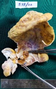
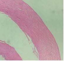
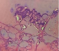
Struma Ovarii is an uncommon variant of ovarian monodermal mature cystic teratoma consisting of thyroid tissue. Preoperative diagnosis of it is difficult as the clinical presentation and ultrasound imaging are often similar to that of ovarian neoplasm. These patients, mostly with benign diseases, often have more extensive surgery than necessary. We present a case report of a 60-year-old patient with a preoperative diagnosis of ovarian neoplasm post-operatively diagnosed as struma ovarii on histopathology which is confirmatory. This case report aims to highlight the importance of associating preoperatively clinical findings and investigations to avoid extensive surgery. When suspected it is crucial to evaluate thyroid status to avoid complications of hyperthyroidism.

Keywords: Monodermal Teratoma, Struma Ovarii, Suspected Ovarian Malignancy, Thyroid Follicles

Corresponding Author How to Cite this Article To Browse
Susheela Gayam, DNB, DGO– HOD & Senior Consultant, Department of Obstetrics and Gynaecology, Vijay Marie Hospital, Hyderabad, Telangana, India.
Email:
Susheela Gayam, Geeta Rani, Laxmi VVS, Trilok, Struma Ovarii – A Case Report. Obs Gyne Review J Obstet Gynecol. 2023;9(1):1-4.
Available From
https://obstetrics.medresearch.in/index.php/joog/article/view/159

Manuscript Received Review Round 1 Review Round 2 Review Round 3 Accepted
2023-01-07 2023-01-09 2023-01-16 2023-01-23 2023-01-30
Conflict of Interest Funding Ethical Approval Plagiarism X-checker Note
Nil Nil Yes 16%

© 2023by Susheela Gayam, Geeta Rani, Laxmi VVS, Trilokand Published by Siddharth Health Research and Social Welfare Society. This is an Open Access article licensed under a Creative Commons Attribution 4.0 International License https://creativecommons.org/licenses/by/4.0/ unported [CC BY 4.0].

Introduction

Struma Ovarii is a rare variant of monodermal ovarian mature cystic teratoma consisting of thyroid tissue [1]. It comprises 1% of all ovarian tumours and 2.7% of all dermoid tumours [2]. It is usually a benign condition; however, malignant transformation occurs in less than 5% of them [3]. It is difficult to diagnose these cases preoperatively as there are no specific clinical, radiological or serum markers in the absence of thyroid abnormality. Most cases are diagnosed on histopathology [4,5]. A typical feature of struma ovarii on doppler is the presence of well-defined solid tissue with a smooth margin that is vascularised - “struma pearl’[6]. Subtle radiological signs and clinical suspicion can avoid extensive staging laparotomies in patients with struma ovarii. The purpose of reporting this case study is to describe a case of struma ovarii and go through a review of the literature to stress the importance of conservative management.

Case Report

60-year-old patient with post-hysterectomy presented with dull aching lower abdominal pain for 3 months with no history of loss of appetite/weight or any symptoms of hyperthyroidism. General and systemic examinations were unremarkable. The thyroid was normal. Abdominal examination showed a transverse scar, no mass was palpable. On per speculum vault appeared normal. On bimanual examination a non-tender, cystic mass of 10x10cm arising from the pelvis was felt in the midline. Ultrasonography displayed a pelvic mass of 9.5x9.1cm with multiple septa and a solid nodule suggestive of ovarian neoplasm. CT pelvis revealed a large well defined thin-walled lobulated cystic mass measuring 11.8x10cm with calcific solid excrescence and few septations within arising from the right adnexa suggestive of ? ovarian neoplasm; no lymphadenopathy. On investigation, TSH 7.62 miu/L, Ca-125 4.8mIU/L, Ca19.9 <3mIU/L, C-Xray were normal. The patient underwent a Laparoscopic right salpingo-oophorectomy. Intraoperative findings included: no free fluid in the abdomen/pelvis, post hysterectomy status, left adnexa not visualised, right ovarian cyst measured 12x12cm with thick yellowish fluid, no normal ovarian tissue, POD and the rest of the viscera were normal.

obsgyne_159_01.JPG

Figure1: Gross Specimen showing thyroid tissue   

obsgyne_159_02.JPG

Figure 2: Low power view - Cyst wall without the Lining Epithelium;


Gross specimen and histopathology showed benign cystic monodermal teratoma-Struma Ovarii with thyroid tissue composed of benign follicles of variable sizes and a lumen filled with abundant colloid. No evidence of atypical/immature neural elements (Figs 1-4).

obsgyne_159_03.JPG

Figure 3: Cyst Wall and Thyroid Tissue

obsgyne_159_04.JPG

Figure 4: Thyroid Tissue

Discussion

Struma ovarii is defined as a mature teratoma comprising thyroid tissue exclusively or predominantly [7]. Most cases are diagnosed on histopathology [5]. It is difficult to diagnose struma ovarii preoperatively as there are no specific clinical, radiological or serum markers in the absence of thyroid abnormality. The incidence of thyroid hyperfunction is reported to be 5-8% in these patients [8]. Often they require extensive staging laparotomy for suspected ovarian malignancy.

Therefore, it is important to correlate and evaluate based on all the clinical findings and investigations preoperatively. Surgery is the primary modality of management for all ovarian tumours but conservative surgery is recommended for struma ovarii and the laparoscopic approach is the preferred route [5]. It can mimic ovarian malignancy clinically, when presented with a complex ovarian mass, with ascites and elevated Ca-125 [9]. Benign struma ovarii has a good prognosis and survival without any significant long-term problems[5].

Conclusion

Although the diagnosis of struma ovarii is difficult preoperatively, correlating all the clinical manifestations, investigations and imaging studies which were all in favour of benign pathology in this case, the conservative approach was undertaken and laparoscopic salpingo-oophorectomy was done instead of subjecting the patient to extensive surgery. A high index of suspicion and relevant preoperative investigations can help to differentiate and distinguish between benign and malignant ovarian tumours preoperatively so that extensive laparotomies can be avoided.

Author Contributions: All authors contributed to drafting the article and approved the final version to be published.

Consent: 'Written Informed Consent' was obtained from the patient for publication of this case report with clinical details and accompanying images.


Reference

01. Yoo SC, Chang KH, Lyu MO, Chang SJ, Ryu HS, Kim HS. Clinical characteristics of struma ovarii. J Gynecol Oncol. 2008 Jun;19(2):135-8. doi: 10.3802/jgo.2008.19.2.135. Epub 2008 Jun 20. PMID: 19471561; [Crossref][PubMed][Google Scholar]

02. Kim SJ, Pak K, Lim HJ, Yun KH, Seong SJ, Kim TJ, et al. Clinical diversity of struma ovarii. Korean J Obstet Gynecol. 2002; 45:748–752. [Crossref][PubMed][Google Scholar]

03. DeSimone CP, Lele SM, Modesitt SC. Malignant struma ovarii: a case report and analysis of


cases reported in the literature with focus on survival and I131 therapy. Gynecol Oncol. 2003 Jun;89(3):543-8. doi: 10.1016/s0090-8258(03)00141-0 [Crossref][PubMed][Google Scholar]

04. Rosenblum NG, LiVolsi VA, Edmonds PR, Mikuta JJ. Malignant struma ovarii. Gynecol Oncol. 1989 Feb;32(2):224-7. doi: 10.1016/s0090-8258(89)80037-x [Crossref][PubMed][Google Scholar]

05. Singh P, Lath N, Shekhar S, Goyal M, Gothwal M, Yadav G, Khera P. Struma Ovarii: A Report of Three Cases and Literature Review. J Midlife Health. 2018 Oct-Dec;9(4):225-229. doi: 10.4103/jmh.JMH_53_18 [Crossref][PubMed][Google Scholar]

06. Savelli L, Testa AC, Timmerman D, Paladini D, Ljungberg O, Valentin L. Imaging of gynecological disease (4): clinical and ultrasound characteristics of struma ovarii. Ultrasound Obstet Gynecol. 2008 Aug;32(2):210-9. doi: 10.1002/uog.5396 [Crossref][PubMed][Google Scholar]

07. Roth LM, Talerman A. The enigma of struma ovarii. Pathology. 2007 Feb;39(1):139-46. doi: 10.1080/00313020601123979 [Crossref][PubMed][Google Scholar]

08. Ayhan A, Yanik F, Tuncer R, Tuncer ZS, Ruacan S. Struma ovarii. Int J Gynaecol Obstet. 1993 Aug;42(2):143-6. doi: 10.1016/0020-7292(93)90628-a [Crossref][PubMed][Google Scholar]

09. Sinha NK. Struma ovarii with elevated ca-125 levels and ascites mimicking advanced ca ovary. J Clin Diagn Res. 2014 Mar;8(3):140-1. doi: 10.7860/JCDR/2014/8005.4138. Epub 2014 Mar 15 [Crossref][PubMed][Google Scholar]