E-ISSN:2455-5444
P-ISSN:2581-4389
RNI:MPENG/2017/74037

Case Report

Abdominal Pregnancy

Obs Gyne Review - Journal of Obstetric and Gynecology

2022 Volume 8 Number 1 January February
Publisherwww.medresearch.in

Abdominal Pregnancy at 38 weeks with good maternal and perinatal outcome

Chaudhary A.1*, Varma U.2, Gupta N.3, Chaudhary D.4, Varma A.5, Shanker J.6
DOI: https://doi.org/10.17511/joog.2022.i01.01

1* Anjali Chaudhary, Senior Consultant, Department of Gynecology and Obstetrics, Aarogya Hospital, Delhi, Delhi, India.

2 Umesh Varma, Senior Consultant, Department of Medicine, Aarogya Hospital, Delhi, Delhi, India.

3 Nishchal Gupta, Consultant, Department of Anesthesia, Aarogya Hospital, Delhi, Delhi, India.

4 Deepali Chaudhary, Consultant, Department of Psychology, Aarogya Hospital, Delhi, Delhi, India.

5 Aditya Varma, Medical Student, , Sri Ramchandra Institute of Higher Education and Research, Chennai, Tamil Nadu, India.

6 Jahnvi Shanker, Intern, , Sri Ramchandra Institute of Higher Education and Research, Chennai, Tamil Nadu, India.

Introduction: Abdominal pregnancy is a very rare form of ectopic pregnancy, associated with high morbidity and mortality for both fetus and mother. An advanced abdominal pregnancy with good fetal and maternal outcomes is therefore a more extraordinary occurrence in the modern developed world. Case: We present a case of abdominal pregnancy which was an emergency diagnosis on OT table while performing elective LSCS for 38 weeks with breech with previous LSCS with normal documented prenatal sonographies (Fig S6, S7). It is extremely rare to first diagnose abdominal pregnancy at the time of elective caesarean section for another indication. Management: Post delivery of a baby by breech extraction, torrential bleed and massive hemorrhage from placenta adhering to bowel and omentum was a big challenge. Provisional differential diagnosis was that of placenta percreta. This led team to follow emergency hemorrhage protocol of laparotomy, total abdominal hysterectomy under blood and blood products coverage with intravascular volume expanders. At the time of laparotomy, the fetus was located in the pelvis covered by the amniotic sac, with distortion of the entire right adnexa and invasion to the right parametrium and the right side pelvic wall. The placenta invaded the pouch of Douglas, the omentum and the bowel on the right pelvic wall. Massive hemorrhage was a challenge and was dealt with successfully (fig 1). Injection methotrexate (50 mg) was used to expedite degeneration of the trophoblastic tissue in the residual placenta. Result: 2.65kg infant (fig S1) and healthy mother were discharged on postoperative 4day.

Keywords: Abdominal pregnancy, Hemorrhage, Laparotomy

Corresponding Author How to Cite this Article To Browse
Anjali Chaudhary, Senior Consultant, Department of Gynecology and Obstetrics, Aarogya Hospital, Delhi, Delhi, India.
Email:
Anjali Chaudhary, Umesh Varma, Nishchal Gupta, Deepali Chaudhary, Aditya Varma, Jahnvi Shanker, Abdominal Pregnancy at 38 weeks with good maternal and perinatal outcome. Obs Gyne Review J Obstet Gynecol. 2022;8(1):1-5.
Available From
https://obstetrics.medresearch.in/index.php/joog/article/view/151

Manuscript Received Review Round 1 Review Round 2 Review Round 3 Accepted
2021-12-06 2021-12-08 2021-12-15 2021-12-22 2021-12-29
Conflict of Interest Funding Ethical Approval Plagiarism X-checker Note
Nil Nil Yes 17%

© 2022by Anjali Chaudhary, Umesh Varma, Nishchal Gupta, Deepali Chaudhary, Aditya Varma, Jahnvi Shankerand Published by Siddharth Health Research and Social Welfare Society. This is an Open Access article licensed under a Creative Commons Attribution 4.0 International License https://creativecommons.org/licenses/by/4.0/ unported [CC BY 4.0].

Introduction

Abdominal pregnancy is a rare type of ectopic pregnancy where the developing embryo implants and grows within the peritoneal cavity [1].  The prevalence of ectopic pregnancy is 1to2% with 95% occurring in the fallopian tube. Abdominal pregnancy account for up to 1.4% of all ectopic pregnancy with an estimated incidence of 1:10,000 live births [2]. The first documented case of abdominal pregnancy was reported in the year 1708. Primary abdominal pregnancy occurs when the fertilized ovum implants directly into the peritoneal cavity. It is a less common type. Secondary abdominal pregnancy occurs when the fertilized ovum implants into the fallopian tube or uterus and then due to tubal abortion or rupture of the fallopian tube or uterus, the fetus comes to live and develop in the mother’s abdominal cavity [3]. In our case, it is unclear if abdominal pregnancy is a result of secondary implantation from an aborted tubal pregnancy or a result of primary implantation from intra-abdominal fertilization. Symptoms of an abdominal pregnancy are very nonspecific and often include abdominal pain, nausea, vomiting, palpable fetal parts, fetal malpresentation, pain on fetal movement and displacement of the cervix [4]. With remarkable advances in radiographic technology, an early discovery of an extrauterine pregnancy should be a practicable endeavour. Associated risks for developing abdominal pregnancy are endometriosis, pelvic inflammatory disease, assisted reproductive techniques, tubal occlusion and multiparty. Most often the pregnancy did not survive and often resulted in extraction of the dead fetus with increased maternal mortality [5]. Abdominal pregnancy is associated with high morbidity and mortality with the risk for death 7 to 8 times greater than from tubal ectopic pregnancy and 50 times greater than from intrauterine pregnancy [5]. In view of the rarity and lack of management guidelines of advanced abdominal pregnancy, we expose this case of abdominal pregnancy to present the symptoms associated that could lead to early recognition and the successful management that resulted in a good maternal and fetal outcome.

Case Report

A 31-year-old G2 P1 L1 at 38 weeks with previous LSCS was planned for elective LSCS under S/A on 9/8/21 in view of breech and 3.9 mm scar thickness

(on USG). The patient reported first to us at 28 weeks for pain abdomen. The patient had right side DJ stenting at 28 weeks gestation for urosepsis due to lower right ureteric obstruction. The patient did not complain of any pain thereafter. Her previous reports and regular ultrasounds were all reported normal. Her first ultrasonography was done at 15 weeks gestation. Her vitals were normal. An abdominal examination revealed generalized tenderness with fetus in breech position and mild scar tenderness, fundal height was term size. FHS was regular. Pelvic examination revealed a long, firm, closed cervix slightly deviated to the patient’s right side. It is extremely rare to first dx abdominal pregnancy at the time of elective caesarean section for another indication. During LSCS, after opening the peritoneum a large sac with full-term live baby in the breech presentation was felt in between the peritoneal sac. It was opened, clear liquor was drained and the baby and a viable female infant were delivered via breech extraction with an Apgar score of 9/9 at 1 and 5 minutes with the weight of 2.65 kg (fig 1). The placenta was adhered to the right uterine wall serosa, posteriorly in the pouch of Douglas, along with the right pelvic wall with omental and bowel attachments (fig 1). A tentative diagnosis of placenta percreta versus concealed placental abruption was made. In view of torrential bleed and massive hemorrhage protocol, the emergency term was alarmed for laparotomy. The decision was made to proceed for hysterectomy and removal of placental tissue due to continuous bleeding after taking consent from attendants and patient herself who was in spinal anesthesia and after that intubated and given general anesthesia. The uterus was exteriorized due to massive bleeding and distortion of anatomy. Total abdominal hysterectomy was performed and excision of most of the placental tissue occupying the right side of pelvic floor adhering to bowel, mesentery and omentum along with multiple feeding vessels which were carefully ligated. Homeostasis was secured some bits of placental tissue was left in situ, which was not bleeding, that was dealt with INJ. Methotrexate (50MG) Intramuscular during the postoperative period. The intra-operatively patient received 4 FFP and 3PRBC. Along with intravascular volume expanders. The estimated blood loss was 1.5litres. The patient was then transferred to ICU for further observation and extubated the following morning. The patient was discharged with the full term healthy female of 2.65 kg on day 4 of surgery.


The baby had mild facial and cranial asymmetry (Fig 1). Mother and baby are doing fine and being followed up closely.

A Pathology report revealed that the placenta weighed 470gms with 10x8x4 cm (fig 3,4). A 15cm segment of tri vessel umbilical cord, 1.2 cm in diameter having eccentric cord insertion. Amniosum nodosum was absent. Placenta was congested with mild chronic placental insufficiency.

The uterus was described as intact and weighed 550gms measuring 15x10x8 cm (Fig 3, 4) with thick endometrium, decidual changes and focal autolysis. No chronic villi or trophoblastic cells were identified in the endometrium. Myometrial hyperplasia was seen.

Discussion

Primary abdominal pregnancy refers to an extrauterine pregnancy where implantation of fertilized ovum occurs directly in the abdominal cavity while secondary abdominal pregnancy is a tubal pregnancy that ruptures with reimplantation within the abdominal cavity usually resulting in tubal or ovarian damage [2]. Extrauterine pregnancy beyond 20 weeks gestation and with a viable fetus is a rare condition [6]. Nunyaluendo and Einterz, in a recent review of 163 cases of abdominal pregnancy, revealed that identification of the condition is often missed with only 45% cases diagnosed during the prenatal period. Abdominal pregnancy can be a little more commonly diagnosed for the first time during laparoscopy or laparotomy performed for tubal ectopic pregnancy but it is extremely rare to first diagnose the abdominal pregnancy at the time of elective caesarean section for some other indication [7, 8]. This is what was there in our case. A high index of suspicion is required to make a diagnosis as both clinical signs and symptoms are not specific. The clinical symptoms of an uncomplicated abdominal pregnancy are persistent abdominal or suprapubic pain (100%) no delay in mensuration, bloody vaginal discharge, gastrointestinal symptoms like nausea and vomiting (70%), painful foetal movements (40%), general malaise, and altered bowel movement. The most common signs reported are abdominal tenderness (100%), abnormal foetal lie (70%), easily palpating the babies part and displaced uterine cervix (40%) [4]. In our patient, the baby was always in the breech

position and abdominal examination was unremarkable as she reported first to us at 28 weeks gestation [9]. Abdominal pregnancy is often associated with foetal deformities such as facial and cranial asymmetry, joint abnormalities limb deformity and central nervous deformity in about 21% of cases [10]. In our case, there was mild facial and cranial asymmetry (Fig 2) [10]. The most common complication of abdominal pregnancy is bleeding from the placental implantation site which can be massive and life-threatening and is the most common cause of maternal morbidity and mortality as high as 30-40% [4, 11]. The massive hemorrhage that mostly occurs with surgery is due to a lack of constriction of blood vessels after placental separations. The placenta can be attached firmly to the parietal peritoneum, mesentery and bowel [12]. Removing the placenta can result in catastrophic hemorrhage and damage to adjacent structures. The decision to remove or leave the placenta should depend on the extent of the placentation particularly with the bowel and omental involvement as well as on the expertise of the surgeon. Because of increased postoperative morbidity and mortality, it is not advisable to leave the placenta in situ [5]. In our case, massive transfusion protocol was applied as per hospital protocol. While if the placenta is left in situ it may lead to secondary hemorrhage, abscess formation, adhesions, coagulopathy, failure of lactogenesis which can require second surgery and also a longer follow up along with other complications. There can be torrential hemorrhage from the placental separation and rapid surgical action is necessary to save the woman’s life [4]. The various manoeuvre can be used to control bleeding that includes compression of bleeding site, ligating the vascular pedicles, lavage with cold saline and local or systemic coagulation promoting agents. The removal of the organ to which the placenta is attached or adherent like hysterectomy, salpingo-oophorectomy, omentectomy can be justified to control the hemorrhage [4,11]. Abdominal packing can be done for uncontrolled hemorrhage.

In this case, the placenta has adhered to the omentum, mesentery and bowel and on the right pelvic wall along with extension into the POD. The decision was made intraoperatively for hysterectomy to obtain adequate haemostasis along with controlling bleeding from ligating vascular pedicles attached to the omentum, mesentery and


bowel. It is very rare to have live advanced abdominal pregnancy reaching term and being successfully managed surgically with a healthy mother and newborn.

obsgyne_151_1.JPG 
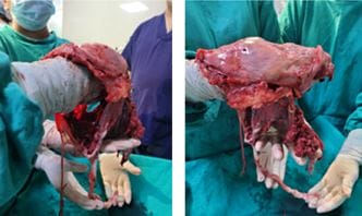
Fig 1: Placental Tissue.

obsgyne_151_02.JPG 
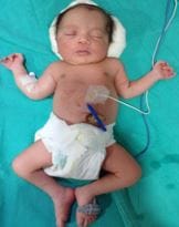
Fig 2: Healty Neonate.

obsgyne_151_03.JPG 
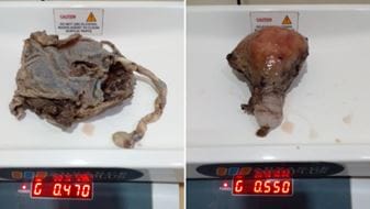
Fig 3: Gravid Uterus & placenta.

obsgyne_151_04.JPG 
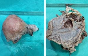
Fig 4: Gravid uterus & placenta.

Conclusion

A high index of suspicion and recognition of signs and symptoms are therefore detrimental to diagnosis and guide to a prompt surgical emergency. Prompt delivery of the fetus, followed by and control of hemorrhage and decision of placenta removal are the greatest challenges. Adequate personnel including anesthesia, pediatricians, and the general surgeon may be necessary for successful management along with adequate blood products facilities available.

Permission from Patient: Authors declare that they have taken written permission from parents for publication of this case report. Journal will not be responsible for any conflict between patient and authors.

Reference

01. N. Golz, D. Kramer, D. Robrecht, and H. Mast. Abdominal pregnancy: case report andreview ofliterature,”Geburtshilfe und Frauenheilkunde, vol. 44, no. 12, pp. 816–818, (1984). View at: PublisherSite | Google Scholar [Crossref][PubMed][Google Scholar]

02. E. L. Nwobodo, “Abdominal pregnancy: a case report. Annals of African Medicine, vol. 3, no.4, pp.185–196, (2004). View at: GoogleScholar [Crossref][PubMed][Google Scholar]

03. L. Badria, Z. Amarin, A. Jaradat, H. Zahawi, A. Gharaibeh, and A. Zobi, Full-term viable abdominal pregnancy: a case report and review." Archives of gynaecology and obstetrics 268.4 (2003): 340-342 [Crossref][PubMed][Google Scholar]

04. Dover, Richard W. , and Martin C. Powell.


Management of a primary abdominal pregnancy. American Journal of Obstetrics & Gynecology 172.5 (1995): 1603-1604 [Crossref][PubMed][Google Scholar]

05. Atrash HK, Friede A, Hogue CJ. Abdominal pregnancy in the United States: frequency and maternal mortality. Obstet Gynecol. 1987 Mar;69(3 Pt 1):333-7. [Crossref][PubMed][Google Scholar]

06. Rahman MS, Al-Suleiman SA, Rahman J, Al-Sibai MH. Advanced abdominal pregnancy--observations in 10 cases. Obstet Gynecol. 1982 Mar;59(3):366-72. [Crossref][PubMed][Google Scholar]

07. Binder, Douglas S. Thirteen-week abdominal pregnancy after hysterectomy. The Journal of emergency medicine 25. 2 (2003): 159-161. [Crossref][PubMed][Google Scholar]

08. Fader, Amanda Nickles, et al. A 14-week abdominal pregnancy after total abdominal hysterectomy. " Obstetrics & Gynecology 109. 2 (2007): 519-521. [Crossref][PubMed][Google Scholar]

09. Baffoe P, Fofie C, Gandau BN. Term abdominal pregnancy with healthy newborn: a case report. Ghana Med J. 2011 Jun;45(2):81-3. doi: 10.4314/gmj.v45i2.68933 [Crossref][PubMed][Google Scholar]

10. Stevens, Cathy A. Malformations and deformations in abdominal pregnancy. American journal of medical genetics 47. 8 (1993): 1189-1195. [Crossref][PubMed][Google Scholar]

11. American College of Obstetricians and Gynecologists. ACOG Practice Bulletin: Clinical Management Guidelines for Obstetrician-Gynecologists Number 76, October 2006: postpartum hemorrhage. Obstet Gynecol. 2006 Oct;108(4):1039-47. doi: 10.1097/00006250-200610000-00046 [Crossref][PubMed][Google Scholar]

12. Varma, R. , L. Mascarenhas, and D. James. Successful outcome of advanced abdominal pregnancy with exclusive omental insertion. Ultrasound in Obstetrics and Gynecology: The Official Journal of the International Society of Ultrasound in Obstetrics and Gynecology 21.2 (2003): 192-194 [Crossref][PubMed][Google Scholar]